How does a pneumothorax look on an X ray?
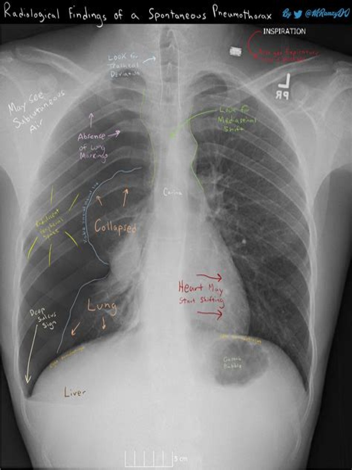
Plain radiograph A pneumothorax is, when looked for, usually easily appreciated on erect chest radiographs. Typically they demonstrate: visible visceral pleural edge is seen as a very thin, sharp white line. no lung markings are seen peripheral to this line.
How does a chest X ray describe a pneumothorax?
A pneumothorax is seen as a region of lucency (dark) around the edge of the lung. This is difficult to see because the lung itself is black too. They are more easily seen on erect chest x-rays as the free air typically rises up to the apex above the lung, making it more visible.
What does a chest xray look like in congestive heart failure?
Radiographic features Chest x-ray findings include pleural effusions, cardiomegaly (enlargement of the cardiac silhouette), Kerley B lines (horizontal lines in the periphery of the lower posterior lung fields), upper lobe pulmonary venous congestion and interstitial edema.
Can a chest xray show a pneumothorax?
A pneumothorax is generally diagnosed using a chest X-ray. In some cases, a computerized tomography (CT) scan may be needed to provide more-detailed images. Ultrasound imaging also may be used to identify a pneumothorax.
What is congenital heart disease chest X-ray?
Congenital heart disease chest x-ray (an approach) A systematic approach to interpreting pediatric chest radiographs is required. A detailed understanding of the normal contours of the cardiomediastinum on chest radiography is essential if abnormalities are to be detected, as well as knowing the range of normal for pulmonary vasculature marking.
How do radiologists identify congenital abnormalities of the thorax?
To accurately identify congenital abnormalities affecting the heart and vessels of the thorax, radiologists must recognize the imaging features and understand their pathophysiologic origin.
What does a box shaped heart mean on a chest xray?
There is often severe right-sided cardiomegaly due to an elongated and enlarged right atrium which may result in an elevated apex. Classically, the heart is described as having a “box shape” on a frontal chest radiograph. 6. Partial Anomalous Pulmonary Venous Return: Scimitar Sign:
What is neonatal pneumothorax?
Andrew Murphy ◉ and Dr Aditya Shetty ◉ et al. Neonatal pneumothorax describes pneumothoraces occurring in neonates. It is a life threatening condition, associated with high morbidity and mortality. The diagnosis is a challenge especially when the amount of air is small and may accumulate along the anterior or medial pleural space.